Ask for a reprint
email :
* Give your email
2020
ACL
|
F.Ren, H.Liu, H.Zhang, Z.Jiang, B.Xia, C.Genevois, T.He, M.Allix, Q.Sun, Z.Li*, M.Gao, 'Engineering NIR-IIb fluorescence of Er-based lanthanide nanoparticles for through-skull targeted imaging and imaging-guided surgery of orthotopic glioma', Nano Today 34 100905 (2020) doi:10.1016/j.nantod.2020.100905
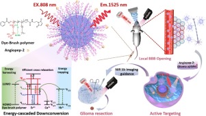
Highly sensitive and specific discrimination of brain tumor margins from the surrounding parenchyma remains a formidable challenge. Limited by the short of photostable probes with deep tissue penetration and high efficiency of crossing the blood-brain-barrier (BBB), the development of fluorescence-guided surgery (FGS) of brain tumors was markedly constrained. Herein, we report the capability of the strong second near-infrared-IIb (NIR IIb, 1500−1700 nm) fluorescence from Er-based lanthanide nanoparticles in imaging-guided surgery of orthotopic glioma. We designed an energy-cascaded Er3+-Ce3+-A3+ (A = Yb, Ho, Tm) system and prepared a series of NaErF4:Ce@NaAF4@NaLuF4 down-conversion nanoparticles (DCNPs) for optimizing the influence of NaAF4 interlayer and Ce3+ dopants. We modified the optimal NaErF4:2.5 %Ce@NaYbF4(0.9 nm)@NaLuF4 DCNPs with Dye-brush polymer (Dye-BP) to facilitate 4I13/2→4I15/2 transition, which leads to an impressive 675-fold enhancement of 1525 nm fluorescence in aqueous solution under 808 nm excitation due to the excellent energy-cascaded downconversion (ECD), in comparison with that of NaErF4 nanoparticles. We further modified these highly bright nanoparticles with tumor-targeting angiopep-2 peptide, and efficiently delivered them to the glioma by using the focused ultrasound sonication (FUS) to temporarily open the BBB. We obtained the highest tumor-to-background ratio (TBR = 12.5) ever reported in the targeted NIR IIb fluorescence imaging of small orthotopic glioma (size < 3 mm, depth> 3 mm) through intact skull and scalp, which was drastically improved to ∼150 after cardiac perfusion and craniotomy to ensure the precise resection of tumor. More importantly, the size of glioma measured from the width of fluorescence profile is very close to that from T2-weighted MRI images. Our work provides the insights into engineering NIR IIb fluorescence of lanthanide nanoparticles, and demonstrates the great potential of NIR IIb fluorescence imaging-guided surgery of tumor.
|
|